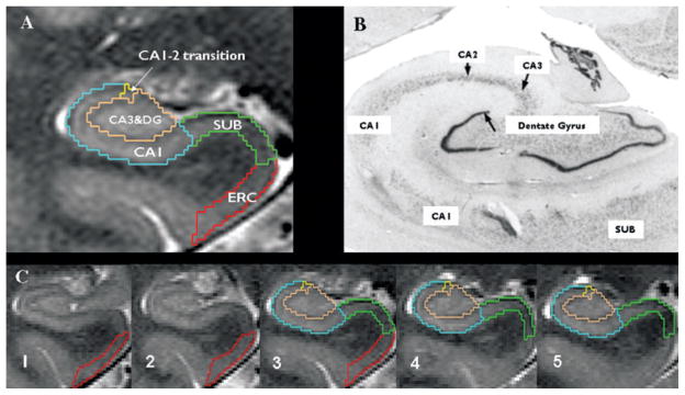
Figure 1.
(A) Parcellation scheme used for manual marking of subfields. Because it is not possible to identify individual hippocampal layers at 4 Tesla, the scheme was based on reliably recognizable anatomic landmarks, even though this resulted in a part of the prosubiculum or pre- and subiculum proper being counted toward the CA1 sector. ERC, entorhinal cortex; CA1-2, CA1-CA2 transition zone (cf Methods in text); CA3&DG, CA3 and dentate gyrus; SUB, subiculum. (B) Histologic preparation of hippocampal subfields. (C) Typical example of hippocampal subfield markings. No. 1 is the most anterior slice in the MRI on which subfields are marked, No. 5 the most posterior slice, and No. 3 (Slice 3), is the referred in the text as “starting” slice. Red, ERC; green subiculum; blue, CA1; yellow; CA1-2 transition; maroon, CA3&DG.
Epilepsia © ILAE

