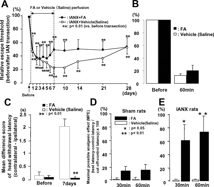
Figure 3.
Head withdrawal threshold to low-intensity mechanical stimulation of the maxillary whisker pad skin (A: continuous i.t. injection of FA, B: i.t. injection of FA at 7 d after IAN transection) and mean differential score of head withdrawal latency to heat stimulation of the whisker pad skin (C: continuous i.t. injection of FA) in awake IANX rats and MPE to high-intensity heat stimulation of the maxillary whisker pad skin in lightly anesthetized sham-control (D) or IANX (E) rats following intrathecal perfusion of FA or vehicle (isotonic saline). The escape threshold to low-intensity mechanical stimulation was significantly reduced after IAN transection compared with that before IAN transection in intrathecal FA- or vehicle (isotonic saline)-perfused rats (A, B). Note that no significant changes in escape threshold to low-intensity mechanical stimulation of the maxillary whisker pad were observed in IANX rats following intrathecal FA perfusion for 7 d (solid circles in A) and intrathecal single injection of FA at 7 d after IAN transection (B). Significant decrease in mean differential score of head withdrawal latency was observed in IANX rats following continuous intrathecal infusion of FA (solid column in C). MPE was significantly increased at 30 and 60 min after intrathecal FA perfusion in IANX rats (solid column in E), but was not changed after vehicle (isotonic saline) administration (open column in E) and FA sham-control rats (D). MPE value was calculated by the following formula: MPE (%) = (test latency − control latency)/(cutoff − control latency) × 100. *p < 0.05, **p < 0.01.

