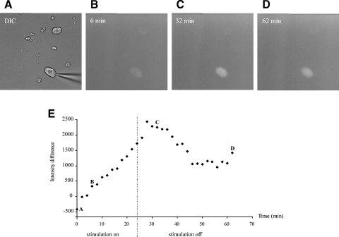
Fig. 1.
Enhanced green fluorescent protein (EGFP) expression in acutely dissociated dorsal root ganglion cells. Dissociated cells were patch clamped and repeatedly depolarized under voltage-clamp conditions while measurements of fluorescence intensity difference were taken at regular intervals. A: multiple cells were included in the field of view, and the difference of intensities were recorded for stimulated and unstimulated cells. B: the fluorescence intensity of the stimulated cell was increased after 6-min stimulation while it was unchanged in the unstimulated cell. C: the fluorescence intensity of the stimulated cell reached a peak even after termination of the stimulation. After a short delay, the intensity decreased. D: the decay of fluorescence intensity appears to be a biphasic decay process, an initial fast phase and a 2nd slower phase. The 2nd phase could take hours to return to baseline. E: plot of fluorescence intensity difference over stimulation time. The stimulation started at time 0 and terminated at time 24 min (- - -). Three time intervals (B–D) were chosen to show the changes in fluorescence intensity in B–D, respectively.

