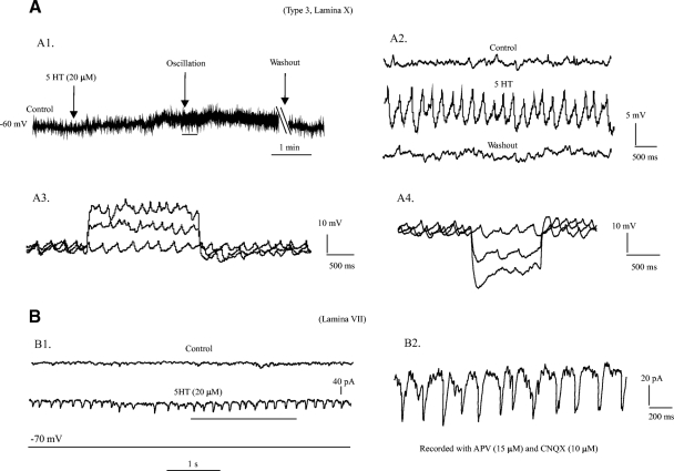
Fig. 7.
5-HT-induced membrane oscillations. A1: the membrane potential was maintained at about −60 mV in a cell of type 3 in lamina X. About 10 mV membrane depolarization was induced by bath application of 20 μM 5-HT. Membrane oscillation was observed 2 min after application of the 5-HT. The 5-HT-induced oscollation could be removed after 15 min washout. A2: the oscillation underlined by black bar in A1 was enlarged to show the details with control and washout. The peak-to-peak amplitude of the oscillation in this cell was ∼7 mV and frequency ∼4 Hz. A3: a 2 s depolarizing step current with step of 50 pA was injected into the same cell. The membrane oscillation remained with a small increase (1–2 Hz) in frequency of the oscillation. A4: a 1-s hyperpolarizing step current with step of −50 pA was injected into the same cell. Again, the membrane oscillation remained but the frequency decrease by ∼2 Hz. The last step evoked sag current and resulted in a membrane depolarization. But the oscillation still remained. B1: 5-HT-induced membrane (current) oscillation was observed in an EGPF neuron in lamina VII with voltage-clamp protocol. The recordings were made with membrane potential clamped at −70 mV and 2-amino-5-phosphonovaleric acid (APV, 15 μM) and 6-cyano-7-nitroquinoxalene-2,3-dione (CNQX, 10 μM) applied to the solution. Top: control; middle: 5-HT-induced oscillation; bottom: clamped voltage. B2: the oscillation underlined by black bar in B1 was enlarged to show the details: peak-to-peak amplitude was ∼50 pA and frequency ∼6 Hz.

