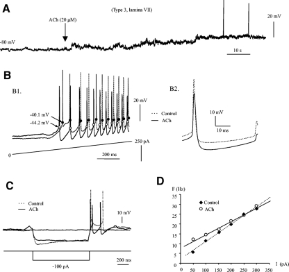
Fig. 9.
ACh modulation of membrane properties of EGFP+ neuron. A: recordings were made from a type 3 cell in lamina VII. A: 20 μM ACh was applied to the bath solution (↓), and the membrane potential was depolarized by ∼17 mV after 1-min application of the drug. Spontaneous firing occurs occasionally during the depolarization of the membrane potential. B1: the hyperpolarization of Vth and increase of afterhyperpolarization (AHP) were shown in the repetitive firings evoked by a 2-s ramp current starting from 0 pA. Spike trains from control (- - -) and ACh (—) were overlapped with unchanged baseline for resting membrane potentials. Voltage thresholds were marked by dots on the spikes (black for control and open for ACh). B2: the 1st 5 spikes from both control and ACh conditions were averaged, respectively, and overlapped on each other (- - -, control; —, Ach) to show the changes in Vth, AHP and action potential (AP) in this neuron. C: a hyperpolarizing current (1 s, −100 pA) was injected into the same cell before and after the ACh application. Membrane potential deflections from the control (- - -) and ACh (—) conditions were overlapped and aligned to the resting membrane potentials. Spikes were elicited from postinhibitory rebound in both conditions. ACh hyperpolarized the Vth by ∼4.1 mV, reduced input resistance by ∼55 MΩ and increased the AHP by ∼3.5 mV in this neuron. D: the F-I curves were plotted for steady-state spikes. ACh reduced the F-I slope by ∼35.6 Hz/nA. The excitability of the cell was increased in the lower range of injected current (≤250 pA) but slightly reduced in the higher range over 250 pA.

