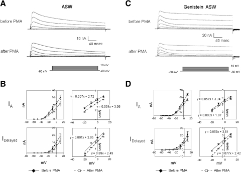
Fig. 9.
Genistein fails to attenuate PMA suppression of K+ currents. A: control preparation exposed to 100 nM PMA showed reductions in IA and IDelayed measured about 20 min following PMA exposure (top traces: currents before PMA; bottom traces: currents ∼20 min after PMA exposure). B: summary data depicting PMA's suppression of K+ currents, for 4 cells, measured 20 min after bath application of PMA. Both the transient (IA) and sustained (IDelayed) components of K+ current were decreased. The voltage dependence of IA and IDelayed was unaffected by PMA. C: genistein-treated preparation exposed to 100 nM PMA also showed reductions in IA and IDelayed measured about 20 min following PMA exposure (top traces: currents before PMA; bottom traces: currents about 20 min after PMA exposure). D: summary data depicting PMA's suppression of K+ currents for cells treated first with genistein, measured 20 min after bath application of PMA. For genistein-treated preparations, the reductions of IA and IDelayed by PMA were comparable to those observed in cells that had not been exposed to genistein (compare with B). Prior to PMA exposure, however, the K+ currents were larger for the genistein-treated cells (compare with A). The voltage dependence of IA and IDelayed was not significantly affected by PMA in the presence of genistein.

