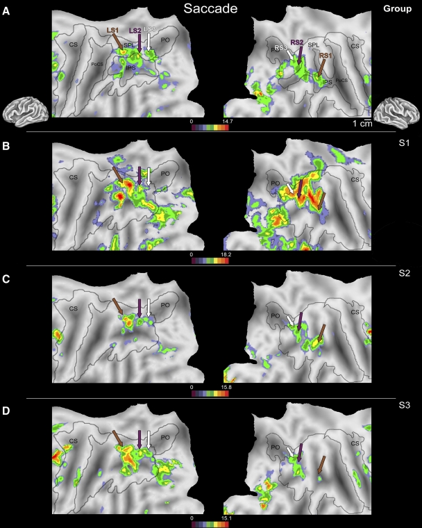
Fig. 1.
Flattened left (left column) and right (right column) hemispheres of a human brain taken from a stereotactic atlas (colin.27), with activity overlays of statistical maps showing areas more active during visually directed saccades to peripheral targets vs. a fixation period from a second-level (random effects) group analysis (A) and from 3 individual subjects (B–D). Brown, purple, and white arrows in B–D indicate locations of peak activations shown in the group analysis in A. Although intersubject variability in activation across medial and caudal cortex can be found in the IPS during visually directed saccades, clusters in the rostral superior parietal lobe of the left hemisphere and rostral IPS of the right hemisphere (LS1/RS1; brown arrow), lateral superior parietal lobe of both hemispheres (LS2/RS2; purple arrow), and the junction of the caudal IPS + PO bilaterally (LS3/RS3; white arrow) were commonly active across the entire group. Variability in the location and extent of activation can be appreciated by comparing the activity around a given arrow across individual subjects (B–D). For example, activation in LS2 (purple arrows) is relatively uniform, with only small shifts in location across subjects. However, the variability in location and extent of activation were slightly greater for LS1 (brown arrows), with somewhat larger shifts in location and amplitude. IPS, intraparietal sulcus; LS1, -2, -3, left saccade activation 1, 2, 3; RS1, -2, -3, right saccade activation 1, 2, 3; PO, parietooccipital sulcus.

