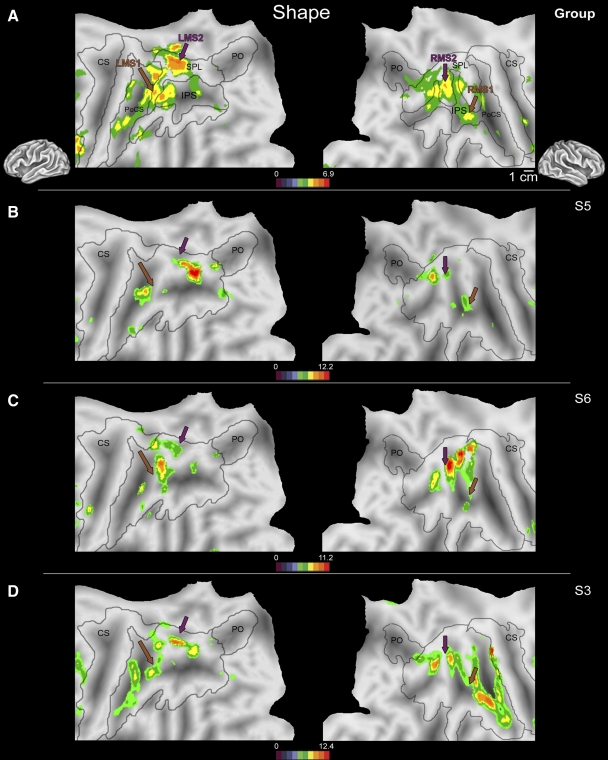
Fig. 3.
Flattened left (left column) and right (right column) hemispheres of a human brain taken from a stereotactic atlas (colin.27), with activity overlays of statistical maps showing areas active during nonvisual manual shape discrimination vs. the shapeless motor control in the second-level (random effects) group analysis (A) and from 3 individual subjects (B–D). Brown and purple arrows indicate locations of activation peaks shown in A. Although intersubject variability in activation across PPC is present during manual shape discrimination at the single-subject level, common clusters of activity at the junction of the IPS + PoCS (LMS1/RMS1, brown arrows) as well as a region of the medial SPL (LMS2/RMS2, purple arrows) are active in all subjects. The region of rostral PPC active during manual shape discrimination in each subject (B–D, brown arrow) was located near area LMS1/RMS1 in the group analysis (A, brown arrow). Likewise, there was a close proximity between the second focus of activity (LMS2/RMS2) in each subject and the location of activity in the group analysis, indicated by the purple arrow in B–D. Conventions as in previous figures. PoCS, postcentral sulcus; LMS1, -2, left manual shape activation; RMS1, -2, right manual shape activation 1, 2.

