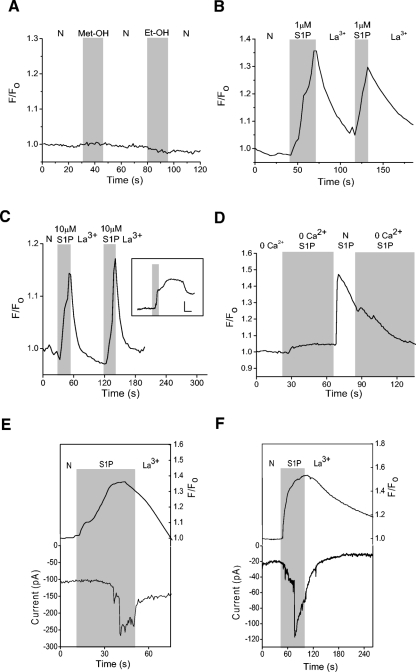
Fig. 8.
S1P generates La3+-sensitive Ca2+ elevations in amacrine cells. Cells were loaded for 1 h with the Ca2+- sensitive dye Oregon Green BAPTA 488. A: a representative example of a fluorescence recording testing the effects of the vehicle for S1P (1% Met-OH) and N,N-dimethyl-d-erythro-sphingosine (DMS, 1% Et-OH), a reagent used in the experiments depicted in Fig. 11. No Ca2+ elevations were seen in response to these compounds. B: a representative recording shows that 1 μM S1P can elicit La3+-sensitive Ca2+ elevations. C: data from a different cell showing a La3+-sensitive cytosolic Ca2+ increase to 10 μM S1P. Inset: data from a different cell showing that in the absence of La3+, the Ca2+ elevations can persist well after the removal of S1P (scale bars are 1.1 F/F0 and 30 s). D: in 0-Ca2+, a small S1P-dependent Ca2+ increase was observed, likely representing release of Ca2+ from internal stores. Re-introduction of external Ca2+ produced a dramatic increase in the S1P-dependent Ca2+ elevation. E: an amacrine cell preloaded with Oregon Green Bapta 488 is voltage-clamped at −70 mV in the perforated-patch configuration. S1P causes a cytosolic Ca2+ increase that precedes the inward S1P-dependent current. F: in a different cell, S1P-induces a cytosolic Ca2+ increase that occurs simultaneously with the S1P-dependent current.

