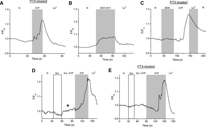
Fig. 9.
The S1P-induced cytosolic Ca2+ increase may be mediated by both S1P1R and S1P3R. A: cells were pretreated overnight (18–22 h) in pertussis toxin (200 ng/ml) to inhibit receptor signaling through Gi. A cell is loaded with Oregon Green BAPTA 488 and then exposed to S1P. S1P still induces a Ca2+ increase despite the pretreatment with PTX. B: to determine if activation of S1P1R alone is sufficient to elicit a Ca2+ increase, SEW2871 (10 μM) is applied. In the presence of SEW2871, a small cytosolic Ca2+ increase is observed. C: in a representative PTX-treated cell, SEW2871 produces little or no Ca2+ elevation. S1P, however, elicited a La3+-sensitive response. D: a cell is exposed to suramin (100 μM) to inhibit signaling through S1P3R. In suramin and S1P, a small cytosolic Ca2+ increase was observed (asterisk). When suramin was removed, the cell responded with a substantially larger Ca2+ increase. E: cells were pretreated overnight in PTX and exposed to suramin to inhibit signaling through Gi and S1P3R. The majority of PTX-treated cells (27/44) showed no S1P-dependent Ca2+ increase in the presence of suramin.

