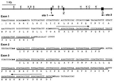
Figure 4.
Partial genomic structure of Oc90. (Upper) Restriction map of the cloned 8.1-kb genomic locus showing the location of three coding exons (solid boxes). Arrows indicate the primers (oto 1 and oto 2) used for RT-PCR amplification of the 5′ Oc90 cDNA. (Lower) sequence of exons 1–3 and flanking intron sequence. Potential splice donor (GT) and splice acceptor (AG) nucleotides are shown in bold. ORFs are indicated with single-letter amino acid notation and those encoding Oc90 are indicated by a solid line. B, BamHI; C, ClaI; E, EcoRI; H, HindIII; K, KpnI, S, SfiI; X, XbaI.

