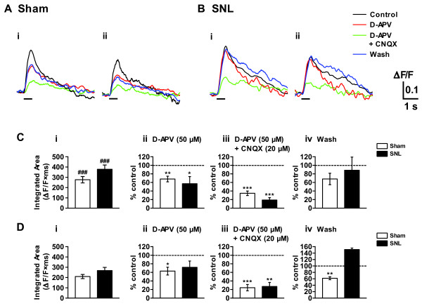
Figure 7.
Primary afferent fiber-evoked spinal excitatory responses in the spinal dorsal horn. (A, B) Optical responses taken before and after the addition of glutamate receptor antagonists (D-APV 50 μM alone or D-APV 50 μM + CNQX 20 μM) in the perfusion solution for 30 min, and also after a wash of the antagonists. The L5 dorsal root (ipsilateral to sham- or SNL-operation) attached to a transverse slice of sham- (A) or SNL- (B) operated spinal cord was stimulated by a suction electrode. Repetitive stimulation with a high intensity (1 mA, 1 ms current pulses, at 20 Hz for 0.5 s as indicated by the bars) was used to activate both A and C fibers. (C, D) Effects of ionotoropic glutamate receptor antagonists on the dorsal root-evoked excitatory responses recorded in the ipsilateral superficial (Ai, Bi, C) and middle (Aii, Bii, D) layers of dorsal horn. Summary of results, testing the effects of an NMDA-receptor antagonist D-APV (Cii, Dii) and mixture of D-APV and a non-NMDA receptor antagonist CNQX (Ciii, Diii) on the optical response elicited by the dorsal root stimulation. Optical responses were also recorded before drug application (Ci, Di) and after washout of the drugs (Civ, Div). The percentage compared to pre-drug response (as 100%) was shown as % control. ###p < 0.001, compared with corresponding middle layer response (Student's t test). n = 16 and 19 for sham and SNL spinal cord slices, respectively. (Cii-Civ, Dii-Div) *p < 0.05, **p < 0.01, ***p < 0.001, compared with pretreatment control (Student's t test). n = 5 for sham and SNL spinal cord slices.

