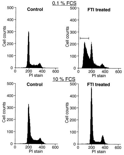
Figure 1.
Flow cytometric analysis of apoptosis in KNRK cells. KNRK cells were plated at a density of 5 × 10⁵ cells/60-mm dish in 10% FCS medium. After 24 h, the medium was changed to either 0.1% FCS medium or 10% FCS medium and the cells were treated with DMSO (Control) or with 20 μM SCH56582 (FTI treated). The cells were collected, stained with PI, and analyzed for DNA content by flow cytometry. Apoptotic cells with a DNA content of <2 N are marked by brackets. At least two separate experiments were carried out with results similar to those shown here.

