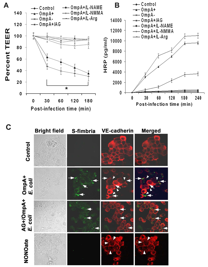
Fig. 1. Effects of NOS inhibitors on OmpA+ E. coli induced permeability of HBMEC monolayers.
Confluent monolayers of HBMEC grown in Transwell inserts were left untreated (Control), pretreated with NOS inhibitors (L-NAME, L-NMMA, or AG) and infected with OmpA+ E. coli or OmpA− E. coli for indicated periods. L-Arginine (L-Arg) was used as a control. TEER (A) and HRP permeability (B) of the monolayers was measured as described in methods section. The experiments were performed at least three times in triplicate and the measurements are means ± SD. The decrease in TEER in HBMEC infected with OmpA+ E. coli or OmpA+ E. coli+L-arginine was significantly lower in comparison to control or OmpA− E. coli infected cells, P<0.001 by Student’s t test. (C) Confluent monolayers of HBMEC in 8 well chamber slides were either uninfected or infected with OmpA+ E. coli for 30 min without or pretreated with AG and subjected to immunocytochemistry. In separate experiments, HBMEC were also treated with NONOate alone. The cells were washed, fixed, permeabilized and stained with anti-VE-cadherin antibodies followed by Cy3 conjugated secondary antibodies. The bacteria were stained with anti-S-fimbria antibody followed by FITC conjugated secondary antibody. The arrows indicate the position of the bacteria and the arrowheads indicate the disassembly of tight junctions. Original magnification × 40.

