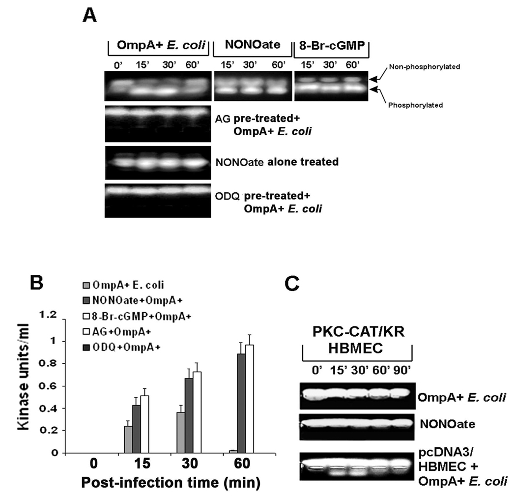
Fig. 10. Activation of PKC-α requires the production of cGMP in HBMEC upon infection with OmpA+ E. coli.
(A) HBMEC were infected with OmpA+ E. coli after pre-treating the cells with AG, NONOate, or ODQ for 1 h as described in the methods section. In some experiments, HBMEC were treated with NONOate or 8-Br-cGMP alone for 1 h. The total cell lysates were subjected to non-radioactive PepTag assay to determine the PKC-α activity. (B) The phosophorylated peptide bands were excised and determined the concentration by a colorimetric assay as described in the methods section and expressed as kinase units/ml. (C) HBMEC were transfected with plasmid alone or a dominant negative form of PKC-α (PKC-CAT/KR), either untreated or treated with NONOate for 1 h, and infected with OmpA+ E. coli for varying periods. The total cell lysates were then subjected to PepTag assay. All experiments were performed at least three times. The error bars in panel B represent standard deviation from the means.

