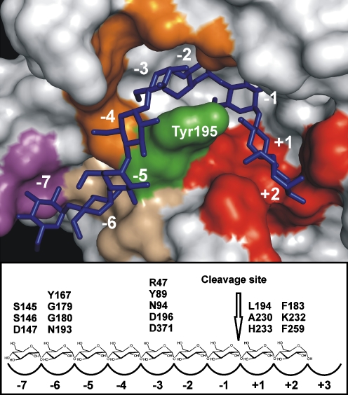
Fig. 3.
Substrate binding at the active site of CGTase. The upper panel shows the binding mode of a maltononaose substrate (blue sticks) at the active site of B. circulans 251 CGTase (crystal structure 1CXK from the protein data bank). Green—Tyr195; red—subsites +1/+2; orange—subsite -3; wheat—subsite -6; and magenta—subsite -7. Figure was created with PyMOL (DeLano 2002). The lower panel gives a schematic overview of the subsites and the residues providing the substrate interactions important for reaction specificity

