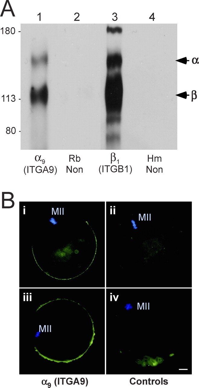
FIG. 1.

Expression of ITGA9 (α9) by mouse eggs. A) Immunoprecipitations from lysates of 300 surface-biotinylated eggs per lane. Lane 1: a purified rabbit anti-ITGA9 polyclonal IgG; lane 2: a nonimmune rabbit IgG (Rb Non); lane 3: an anti-ITGB1 monoclonal antibody (Hmβ1-1); lane 4: a nonimmune Armenian hamster IgG (Hm Non). Sizes shown are Mr × 10−3. B) Immunofluorescence studies of mouse eggs. Panels i and iii: anti-ITGA9 chicken IgY; panel ii: nonimmune chicken IgY; panel iv: anti-ITGA9 chicken IgY preincubated with a 5-fold molar excess of ITGA9 fusion protein. An FITC-conjugated goat anti-chicken IgY secondary antibody was used in all panels, shown in green. Blue staining shows DAPI labeling of the metaphase II DNA (MII). Bar = 10 μm.
