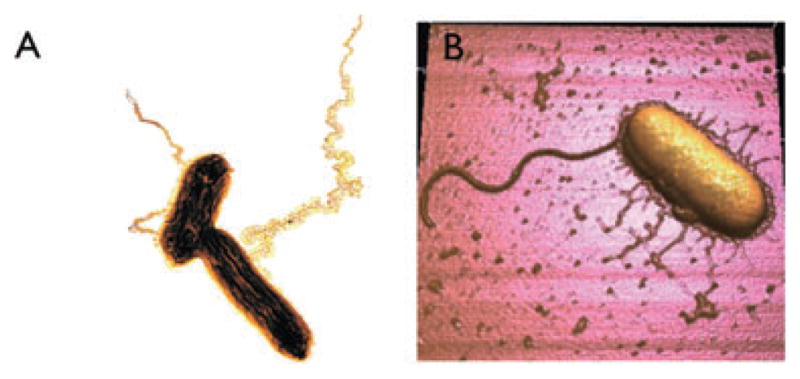
Fig. 2.

‘Nanowires’ produced by two different Fe(III)-reducing bacteria.
A. Transmission electron micrograph of platinum-shadowed ‘geopili’ produced by Geobacter sulfurreducens (courtesy of Gemma Reguera, Michigan State University). The length of the longer cell is approximately 2 μm. The image has been colorized to enhance contrast.
B. Atomic force micrograph of Shewanella oneidensis (courtesy of Pamela Gross, University of Southern California). The large appendage is the flagellum; the shorter pili are electrically conductive nanowires. The length of the cell is approximately 3 μm.
