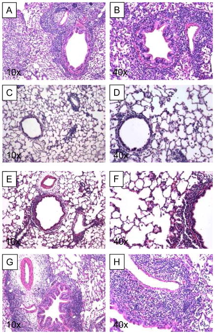
Figure 1. H & E staining of lung after left lung transplantation and adoptive transfer of col(V)-specific effector and regulator T cells.
A and B: 7 days after transfer of 1×10⁷ effector cells into rats subjected to lung isograft transplantation. C and D: 7 days after adoptive transfer of 1×10⁷ effector cells together with 4×10⁷ regulator cells into rats subjected to lung isograft transplantation. E and F: 14 days after adoptive transfer of 1×10⁷ effector cells together with 4×10⁷ regulator cells into rats subjected to lung isograft transplantation. G and H: 7 days after adoptive transfer of 1×10⁷ effector cells together with 4×10⁷ CD4+-T-cell-depleted regulator cells. All slides are H&E stained 5 μm sections of paraformaldehyde fixed lungs.

