
Figure 7. Involvement of Gα subunit (GPA1) in flg22 inhibition of guard cell K+ currents.
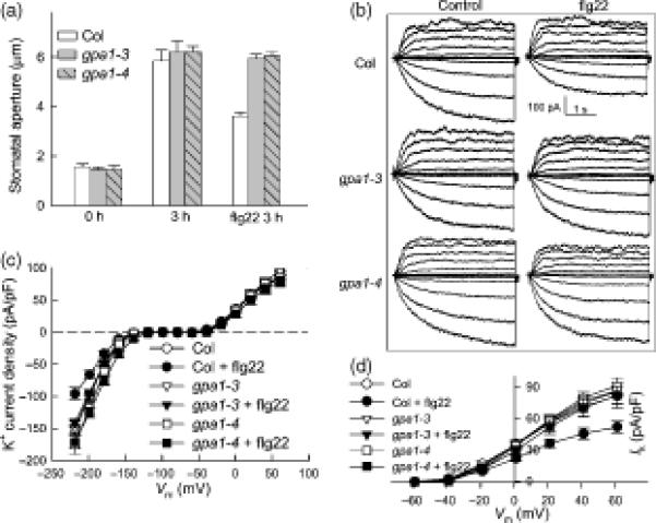
(A) Stomatal apertures of Col-0, gpa1-3 and gpa1-4 plants before light treatment (0 hr) and after 3 hr in the light with or without 5 μM flg22 treatment. n = 3 experiments, with n>110 apertures measured per experiment.
(B) Typical whole-cell recordings at 10 min of Col-0, gpa1-3 and gpa1-4 guard cell K+ currents in the absence or presence of 5 μM flg22. Time and current scales are indicated.
(C) Average current-voltage relationship (mean ± SE) of time-activated whole-cell K+ currents from cells recorded as in (B). n = 22 (Col-0); 17 (Col-0 + flg22); 31 (gpa1-3); 47 (gpa1-3 + flg22); 16 (gpa1-4) and 26 (gpa1-4 + flg22).
(D) Average current-voltage relationship (mean ± SE) of whole-cell outward K+ currents at 20 min from the cells recorded as in B. n = 22 (Col-0); 17 (Col-0 + flg22); 31 (gpa1-3); 47 (gpa1-3 + flg22); 16 (gpa1-4) and 26 (gpa1-4 + flg22).