
Figure 1.
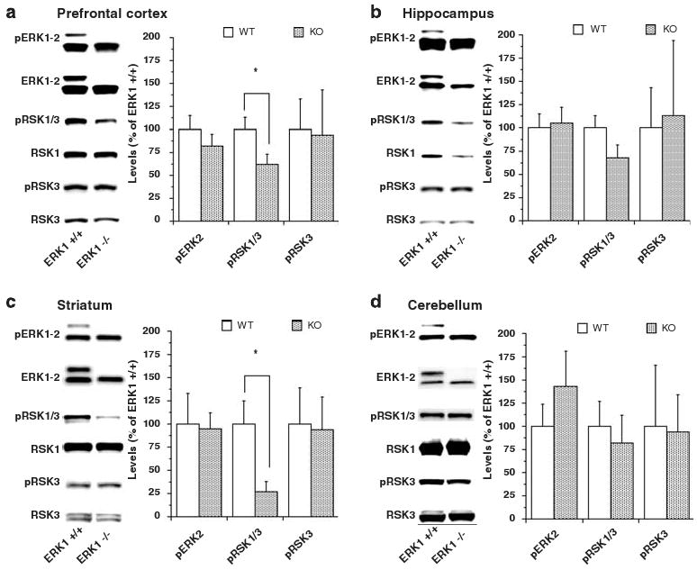
Levels of activated phospho-ERK1/2 (pERK1/2), activated phospho-RSK1/3 (pRSK1/3), activated phospho-RSK3 (pRSK3), ERK1/2, RSK1 and RSK3 in prefrontal cortex (a) hippocampus (b) striatum and (c) cerebellum (d) of wild-type (WT) and ERK1 knockout (KO) mice. Brain samples were extracted on ice, immediately frozen on dry ice and then stored at −80 °C until further analysis. The samples were prepared in a buffer containing protease inhibitor, phosphatase inhibitor I and II cocktails from Sigma. An equal amount of protein in all samples was loaded onto the gel and antibodies indicated in the figure were used according to the manufacturer's recommendations. Densitometric data are presented in the bar graphs as means ± s.e. ERK1 KO mice showed significant reductions in levels of pRSK1/3 in prefrontal cortex (t(35) = 2.07; P= 0.0455) and striatum (t(18) = 2.57; P= 0.0193). Hippocampal levels of pRSK1/3 did not significantly differ between WT and KO mice (t(36) = 1.655; P= 0.1067). *P < 0.05.