
Fig. 3.
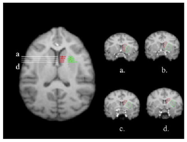
Transaxial view of the chimpanzees with corresponding lines to indicate regions along the anterior–posterior axis representing the caudate (red) and putamen (green). From a to d, coronal views of the tracing of the caudate (refs) and putamen (green) on different slices along the anterior–posterior gradient (a–d). The caudate is outlined in the left hemisphere.