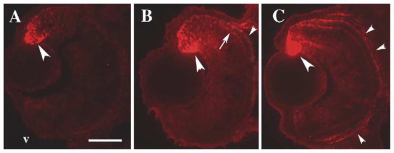
Fig. 5.

Sectioned zebrafish embryo eyes processed for immunocytochemistry with an anti-RALDH2 (dorsal retinal RA synthesizing enzyme) antibody (red fluorescence). Embryos were fixed at 45 hpf (A), 52 hpf (B), and 80 hpf (C). The antibody labels neuroepithelial cells (large arrowheads) and a few developing neurons, including photoreceptors (arrows), in dorsal retina. At 52 and at 80 hpf, some staining is also evident in ventral retina, throughout the RPE (small arrowheads), and in extraocular locations. v, ventral (in all panels); scale bar = 40 μm.
