
Fig. 2. Co-localization of PV, SOM, CR, NPY and VIP with GFP-expressing GABAergic neurons in GAD-GFP transgenic mouse cortex.
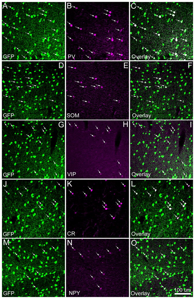
As shown in A–C, essentially all the cells immunopositive for parvalbumin (PV) were co-localized with GFP expressing GABAergic cells in the transgenic mouse in which basically all the GABAergic cells express GFP. The arrows in A–C point to the PV-immunopositive cells overlapping with GFP expression. Similarly, D–F, G–I, J–L and M–O show that nearly all the cells immunopositive for somatostatin (SOM), calretinin (CR), neuropeptide tyrosine (NPY) and vasoactive intestinal peptides (VIP), respectively, were also co-localized with GFP expressing GABAergic cells. The scale bar in O applies to all panels.