
Fig. 4. Double immunochemical staining of mouse cortical sections with different combinations of somatostatin (SOM), calretinin (CR), neuropeptide tyrosine (NPY) and vasoactive intestinal peptide (VIP).
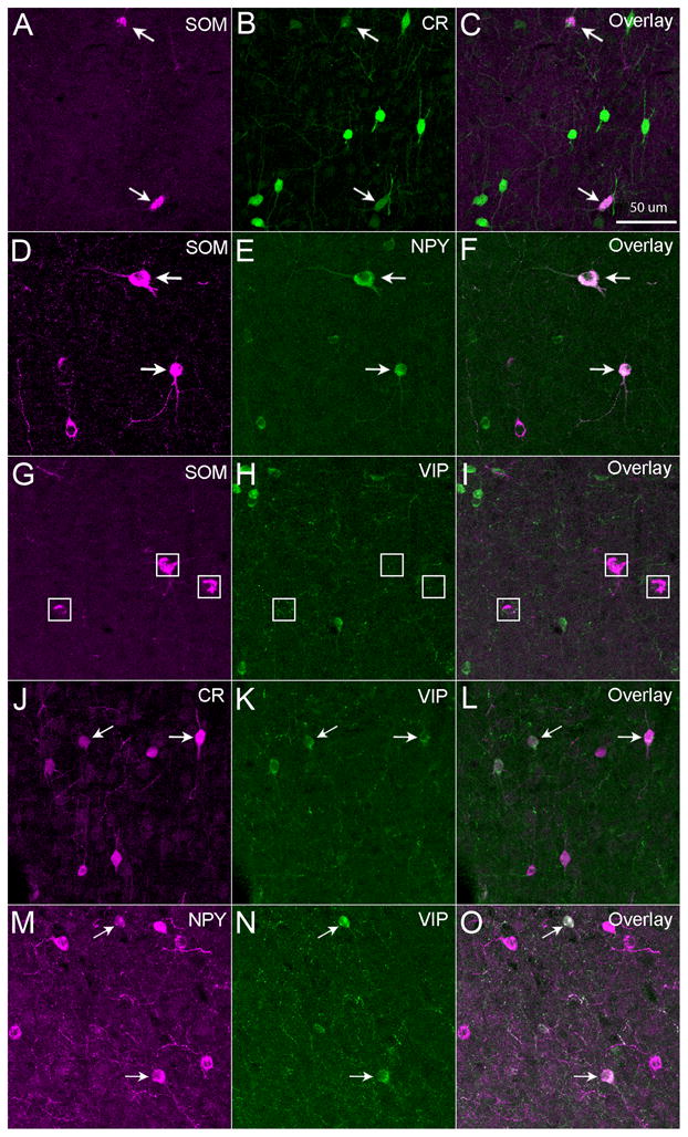
Some SOM+ neurons were immunopositive for CR (A–C) or NPY (D–F), but SOM+ neurons were not immunopositive for VIP (G–I). Some CR+ cells were immunopositive for VIP (J–L), and some NPY+ cells were also immunopositive for VIP (M–O). Arrows point to double-labeled cells. White boxes in panels G–I denote locations of SOM+ cells. The sections were from mouse S1 cortex. The scale bar in C applies to all panels.