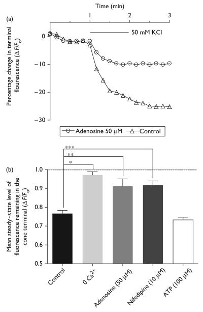
Fig. 3.
Kinetics of Synaptored-C2 destaining in cone photoreceptor terminals with the application of elevated [K+]o superfusate (50 mM). Plot of Synaptored-C2 destaining from a cone terminal treated with elevated [K+]o (triangles) for 1 min and elevated [K+]o for 1 min in the presence of adenosine (50 μM; circles). (b) Adenosine (50 μM) partially inhibited the destaining of Synaptored-C2 fluorescence in synaptic terminals (**P = 0.0014). 0 Ca2+ superfusate essentially prevented destaining (***P < 0.0001). Nifedipine (10 μM) partially inhibited destaining (*P = 0.0003), confirming the role of L-type channels in transmitter release from cones. ATP (100 μM) had no effect on destaining profile (for steady-state fluorescence levels in cones, see text). *P <0.05 compared with an unstimulated normalized total fluorescence value of 1.0.

