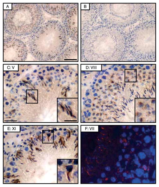
Figure 1.
A study to assess the cellular localization of 14-3-3 in the seminiferous epithelium of adult rat testes. (A–E) Immunohisto-chemical localization of 14-3-3θ in normal adult rat testes, in which frozen cross sections (~7 μm thick) were immunostained using an anti-14-3-3θ IgG (Santa Cruz Biotechnology, Cat. Sc-732; Lot. A2908) (A and C–E) or normal rabbit IgG (B). Low magnification of testes stained with anti-14-3-3θ polyclonal antibody (A) or normal rabbit IgG (B) illustrating the presence of 14-3-3θ in the seminiferous epithelium. In higher magnification, intense staining is obvious surrounding the heads of elongating spermatids at the apical ES site (C and E) as well as round spermatids (D) and at the basal compartment consistent with its presence at the BTB. Bar in (A)=100 μm, which applies to (B), bar in (C)=30 μm, which applies to (D and E), bar in inset in (C)=15 μm, which applies to insets in (D and E) and (F). A study by immunofluorescence microscopy further illustrates the localization of 14-3-3θ in seminiferous epithelium of normal adult rat testes, by using frozen testis cross sections (~7 μm thick) stained with anti-14-3-3θ IgG. CY3-conjugated donkey anti-rabbit secondary antibody (red fluorescence) was used for visualizing 14-3-3θ, confirming the existence of 14-3-3θ at the apical ES in the seminiferous epithelium in a stage VII tubule. Cell nuclei were stained by DAPI. Bar in (F)=12 μm. The Roman numeral after C, D, E and F represents the seminiferous epithelial stage of the tubule.

