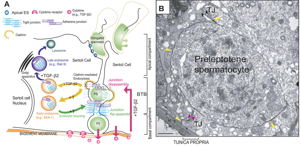
Figure 7.
A model to illustrate that the timely opening and closing (or restructuring) of the BTB to facilitate preleptotene/leptotene spermatocyte migration across the BTB is mediated by the differential effects of T and cytokines on the endocytosis, recycling, and intracellular degradation (or transcytosis) of integral membrane proteins. A) As shown in this schematic drawing, the BTB physically divides the seminiferous epithelium into the basal and apical (or adluminal) compartments. Spermatogonia (type B) differentiate into preleptotene/leptotene spermatocytes (PS) that must traverse the BTB (PSi, preleptotene/leptotene spermatocyte in transit) at stage VIII of the seminiferous epithelial cycle of spermatogenesis. T and cytokines (e.g., TGF-β2 released by Sertoli and/or migrating spermatocytes into the microenvironment of the seminiferous epithelium) (short pink arrow) accelerate the kinetics of internalization of BTB integral membrane proteins via the clathrin-mediated endocytosis pathway (yellow arrows). This disrupts the BTB on the apical end of a migrating PSi at stage VIII of the epithelial cycle to facilitate the transit of PS across the BTB by lowering the steady-state levels of integral membrane proteins locally. Internalized proteins (e.g., occludin) are either targeted to early endosomes via their attachment with an endosome-associated marker protein (e.g., EEA-1) (purple arrow) or sorting endosomes. TGF-β2 perturbs the BTB (long pink arrow) by targeting internalized proteins (e.g., occludin) into late endosomes (e.g., Rab 9) (purple arrow), for their degradation in lysosomes, thereby lowering the steady-state integral membrane proteins at the BTB. T, in contrast, enhances the kinetics of recycling of BTB proteins back to the Sertoli cell surface, perhaps to the basal region of the migration of PS (i.e., PSi) via transcytosis to reassemble the BTB (green arrow). The net result of these interactions thus maintains the immunological barrier in the seminiferous epithelium during the transit of PS across the BTB. B) Electron micrograph of a preleptotene spermatocyte in transit corresponding to the PSi shown in A. This preleptotene spermatocyte was shown to be trapped between the TJ fibrils, as illustrated in this lanthanum study. The TJ fibrils at the apical end of this preleptotene spermatocyte appeared to become leaky (opposing black arrowheads) or opened, as lanthanum (yellow arrowheads) was able to reach the TJ fibrils (yellow arrowhead near the opposing black arrowheads) in this microenvironment to facilitate the transit of this preleptotene spermatocyte. However, new TJ fibrils were detected at the basal portion of this spermatocyte in transit (opposing pink arrowheads) that limited further entry of lanthanum (yellow arrowhead below the pink arrowhead) into the epithelium. Asterisks mark the basement membrane (also known as basal lamina), which is a modified form of extracellular matrix (51); M, myoid cell layer; Nu, Sertoli cell nucleus. Scale bar = 1 µm.

