
Figure 5.
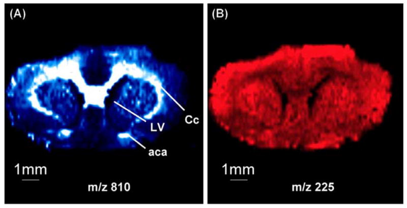
Negative ion DESI MS images of (A) m/z 810 and (B) m/z 255 from a 4 μm coronal section of rat brain tissue illustrate the utility of the method to resolve morphological features in tissues. (Reprinted with permission from reference (Ifa et al. 2007).)