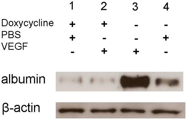
Figure 2. Induced expression of sVEGFR1Fc blocks VEGF-induced breakdown of the blood-retinal barrier.

Tet/IRBP/sVEGFR1Fc mice were given 2 mg/ml of doxycycline in their drinking water or given normal water for 2 weeks and then 1 μl of PBS or PBS containing 10−6 M VEGF was injected into the vitreous cavity. Six hours after injection the mice were perfused with PBS and then the retinas were isolated and retinal homogenates were run in immunoblots using an anti-albumin antibody. The blot was stripped and incubated with anti-β-actin. Retinas from doxycycline-treated mice showed a faint band for albumin whether they received an intraocular injection of PBS (lane 1) or VEGF (lane 2). Mice that were not given doxycycline showed a very strong band for albumin when they received an intraocular Injection of VEGF (lane 3) and a weak band when given an intraocular injection of PBS (lane 4). These results are representative of 3 different experiments.
