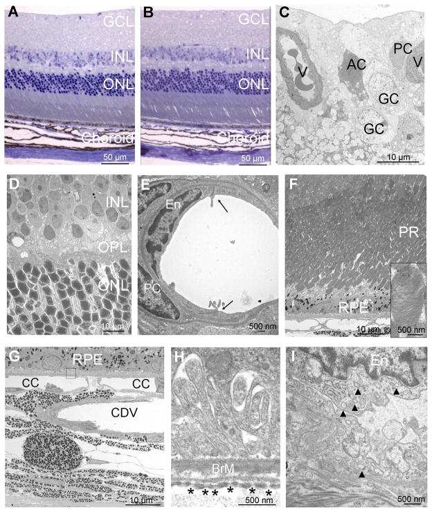
Figure 6. Long term blockade of VEGF does not cause identifiable morphologic changes in the retina, retinal pigmented epithelium (RPE), or choriocapillaris.
Tet/IRBP/sVEGFR1Fc mice or littermate controls were given 2 mg/ml of doxycycline in their drinking water for 7 months. One micron ocular sections stained with toluidine blue appeared normal in both Tet/IRBP/sVEGFR1Fc (A) and control (B) mice. Electron micrographs of the retina, RPE, or choroid of Tet/IRBP/sVEGFR1Fc mice are shown in C–I.
(C) Along the inner surface of the retina, there were normal vessels (V) with normal pericytes (PC), ganglion cells (GC) and astrocytes (AC).
(D) The inner nuclear layer (INL), outer plexiform layer (OPL), and outer nuclear layer (ONL) appeared normal.
(E) High magnification views of retinal vessels showed tight junctions (arrowheads) between normal endothelial cells (EC) surrounded by normal pericytes (PC).
(F) The photoreceptors (PR) and RPE appeared normal and high magnification showed normally stacked disks in photoreceptor outer segments (inset).
(G) The RPE, choriocapillaris (CC) and large choroidal vessels (CDV) appeared normal.
(H) High magnification of the boxed area in (G) shows normal Bruch’s membrane (BrM) with numerous fenestrations in the endothelial cells of the choriocapillaris (asterisks).
(I) High magnification of a choroidal endothelial cell (En) shows numerous vesicles (arrowheads) in the cytoplasm indicative of active vesicular transport. The illustrations are representative of 6 sections from 3 Tet/IRBP/sVEGFR1Fc mice that were examined. The ultrastructure of control mice appeared identical and is not illustrated.

