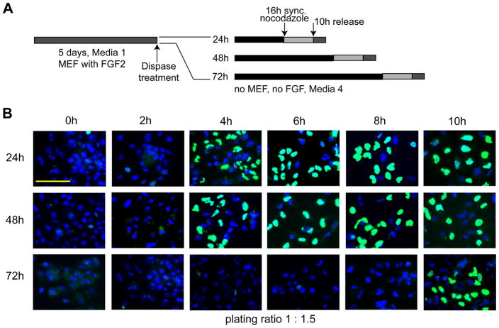
Figure 2. Extension of the G1 phase in human ES cells during early lineage-programming.
(A) Experimental design. (B) Human ES cells were cultured in differentiating media for 24 h, 48 h, and 72 h under feeder-free conditions (first, second and third rows, respectively). Cell cultures were then synchronized using nocodazole for an additional 16 h and subsequently released. BrdU incorporation in nuclei was detected by immunofluorescent microscopy (scale bar = 100 μm) using cells taken at the time of release (0 h) and at 2 h intervals for up to 10 h (first through sixth column, respectively). Cells were plated at a 1:1.5 split ratio (see Figure 3).

