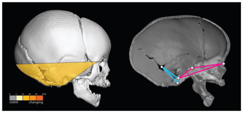
Fig. 4.

Linear distance pairs within the cranial base show significantly reduced integration in the post-operative ISS sample (left). Relative to the pre-operative ISS sample, the postoperative sample shows a marked reduction in the magnitude of morphological integration for linear distance pairs that include a measure from that portion of the cranial base anterior to foramen magnum (shown in pink) and a measure from the inferior portion of the posterior cranial fossa (shown in blue).
