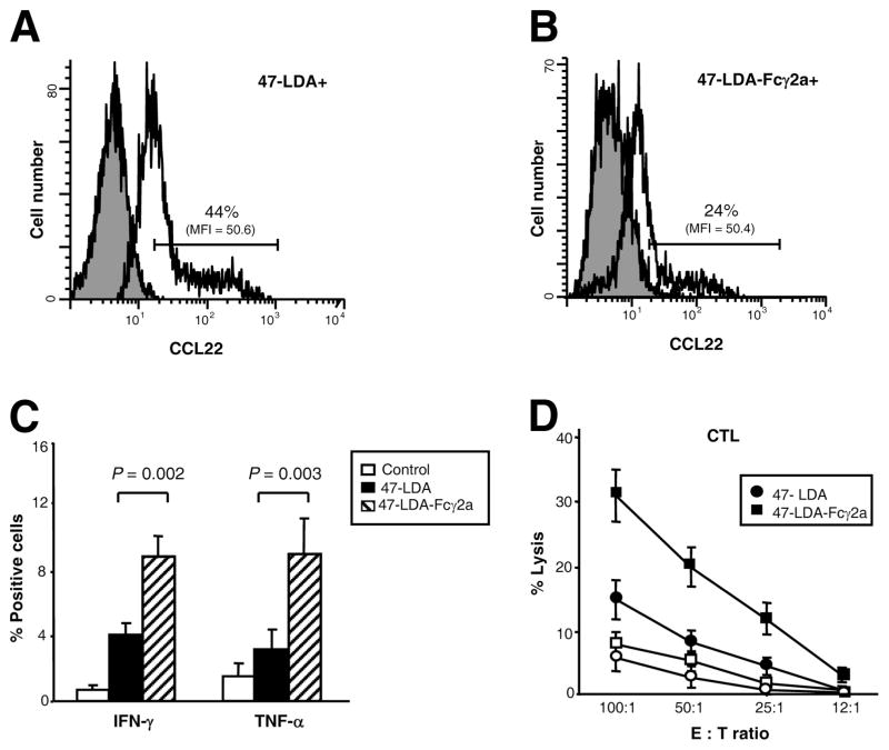
FIGURE 3.
Differential expression of CCL22 in 47-LDA+ and 47-LDA-Fcγ2a+ DCs after LPS stimulation and induction of IFN-γ and TNF-α in CD4+ splenocytes by 47-LDA- and 47-LDA-Fcγ2a-DC vaccines. A and B, Immature DCs were stained with biotinylated 47-LDA polypeptide or 47-LDA-Fcγ2a fusion protein followed by streptavidin-PE. The 47-LDA+ (A) and 47-LDA-Fcγ2a+ (B) DCs were sorted on BD FAC-SAria flow cytometer, incubated with 1 μg/ml LPS for 24 h, and analyzed for CCL22 expression by intracellular staining with rat anti-mouse CCL22 mAb followed by goat anti-rat secondary Ab. The mean fluorescence intensity (MFI) and percentage of cells positive for CCL22 expression are indicated. The gray area denotes background staining assessed using an isotype control Ab. Data are from one representative experiment of three performed. C, Expression of IFN-γ and TNF-α in splenocytes of mice immunized with 47-LDA-DC and 47-LDA-Fcγ2a-DC vaccines (filled and hatched bars, respectively). A/J mice (n = 5) were immunized three times with DCs coated with 47-LDA polypeptide or 47-LDA-Fcγ2a fusion protein after LPS-induced maturation in the presence of IL-15 and IL-21 vectors. Cells isolated from mice immunized with LPS-treated DC served as controls (open bars). Three weeks after the last immunization, the expression of IFN-γ and TNF-α in CD4+ splenocytes was analyzed by intracellular staining after overnight stimulation with DCs expressing the 47-LDA mimotope. D, NXS2 neuroblastoma-specific CTL responses. CD8+ splenocytes from mice immunized with 47-LDA-DC (●) and 47-LDA-Fcγ2a-DC (■) vaccines were obtained by negative selection. Cells were cultured with 47-LDA-expressing DCs (filled symbols) or sham plasmid-transfected DCs (open symbols) at the 20:1 ratio as described in the Materials and Methods section. The CTL activities against NXS2 cells were analyzed in a standard 51Cr-release assay. All determinations were made in triplicate samples, and the SD was <10%. Results are presented as the means ± SD of four independent experiments.

