
Figure 8.
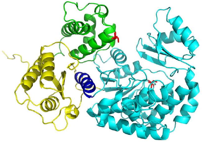
PatchDock docking model for AT3 with linkers (truncated from PDB ID 2QO3, [15]) and DEBS ACP3 (homology model generated by I-TASSER). AT domain, linkers, C-terminal helix and ACP domain are shown in cyan, yellow, dark blue and green respectively. Active site Ser residues are shown as red.