
Figure 3.
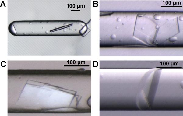
Microphotographs of crystals of enoyl-CoA hydratase from Mycobacterium tuberculosis obtained from SlipChip-based FID. A) A crystal obtained from reagent 15, 20% (w/v) PEG-3000 in imidazole buffer, pH 8.0; B) A crystal obtained from reagent 41, 45% (w/v) PEG-3000 in 0.1 M CHES buffer, pH 9.5; C) A crystal obtained from reagent 8, 2.8 M (NH4)2SO4 in 0.1 M citrate buffer, pH 5.5; D) A crystal obtained from reagent 14, 1.4 M sodium citrate in 0.1M cacodylate buffer, pH 6.5. Reagent numbers correspond to numbering in Table S1.