
Figure 7.
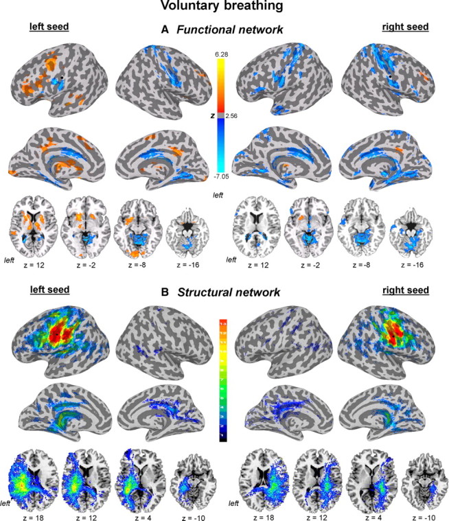
Functional and structural networks of the laryngeal motor cortex associated with voluntary breathing. Functional connections and probabilistic tractography from both left and right hemispheric seed regions (marked as black circle) are presented on the inflated cortical surfaces; subcortical and cerebellar connections are shown on the series of axial images of a single subject in the standard space (p ≤ 0.05, corrected). For functional networks, color scale bar represents z-values, which reflect the strength of PPI correlations, ranging from positive (red-yellow) to negative (light blue-dark blue). For structural networks, color scale bar illustrates probabilistic distribution of structural connections (e.g., the chance of probability of a pathway passing through a given brain region) ranging from 1 to 13 subjects.