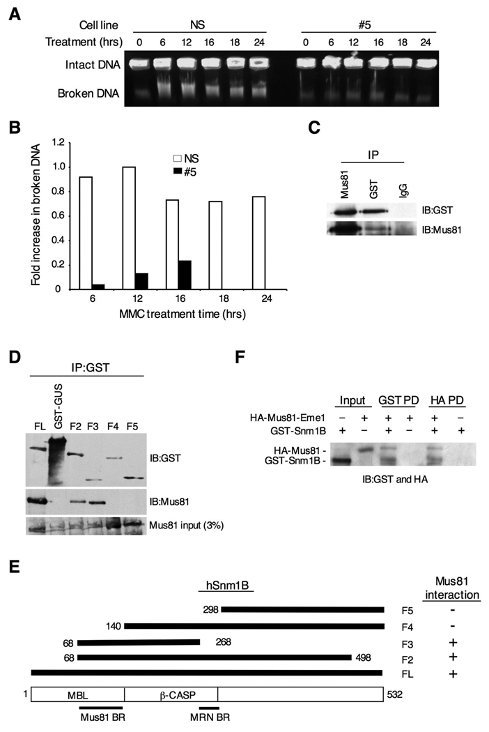
Figure 6. Snm1B is required for fork collapse after MMC treatment.
(A) Double-strand break formation was analyzed by pulsed field gel electrophoresis. Intact DNA stays in the well while broken DNA migrates into the agarose gel. The NS and #5 clones were incubated with MMC (1 ug/ml) for the indicated times, and cells were collected in agarose plugs for gel electrophoresis. (B) Quantitation of results shown in (A). Images were quantitated using ImageJ 1.37v software (developed by Wayne Rasband, NIH). The background present at the 0 hr time point was subtracted from the other time points, and the band with the highest intensity was normalized to a value of 1.0. (C) Snm1B co-IPs with Mus81. GST-Snm1B was transiently expressed in HEK293 cells, and the indicated co-IP assays were performed from lysates. (D) The Mus81 interaction domain maps to the metallo-β-lactamase domain of Snm1B. The indicated GST-SNM1B deletion constructs were expressed in HEK293 cells and the indicated co-IP assays were performed and analyzed by immunoblotting. (E) Schematic depicting the results from (D). (F) Snm1B directly interacts with Mus81-Eme1. Immunoblots showing results of reciprocal pull-down (PD) assays between GST-Snm1B and HA-Mus81-Eme1. HA indicates the hemagglutinin tag.

