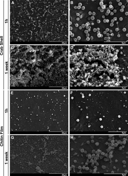
FIG. 1.
F. novicida biofilm formation on chitin surfaces. Images display SEM visualization of F. novicida colonization of crab shell pieces (A to D) and synthetic chitin films (E to H). Individual attached bacteria and small attached microcolonies were observed on crab shell pieces at 1 h (A and B). After 1 week, typical 3D biofilm architecture was observed, consisting of bacteria surrounded by an EPS matrix (C and D). Similar results were obtained after 1 h (E and F) and 1 week (G and H) on synthetic chitin.

