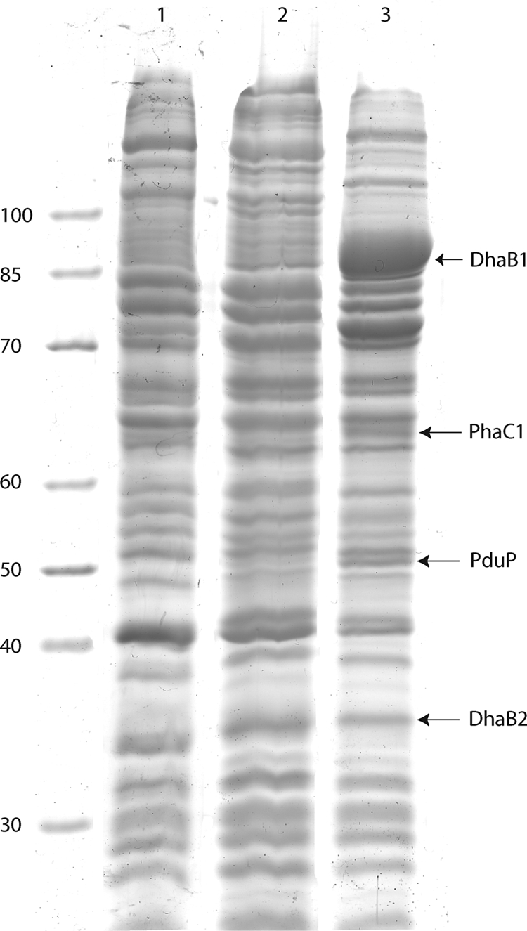
FIG. 3.

Proteins in crude extracts of cells of E. coli HMS174(DE3), E. coli HMS174(DE3)/pCOLADuet-1, and E. coli HMS174(DE3)/pCOLADuet-1::dhaB1B2::pduP::phaC1 grown in LB medium. Protein expression was induced with 1 mM IPTG at an optical density at 600 nm of 0.5, and the cells were then cultivated for an additional 8 h. The proteins were separated using 12.5% (wt/vol) SDS-polyacrylamide gels and stained with Coomassie brilliant blue. Lane 1, E. coli HMS174(DE3); lane 2, E. coli HMS174(DE3)/pCOLADuet-1; lane 3, E. coli HMS174(DE3)/pCOLADuet-1::dhaB1B2::pduP::phaC1. The values on the left indicate molecular mass (in kDa).
