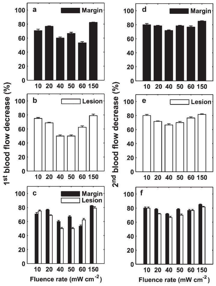
Fig. 7.
Percentage of 1st (a–c) and 2nd (d–f) blood flow decrease relative to the initial value versus fluence rate for lesion and margin regions. The error bars indicate the uncertainties in the estimate of the percentage blood flow decrease, calculated using the criterion introduced in Figure 4a. Generally, the blood flow reduction decreases with an increase of irradiance from 10 to 40 or 60 mW cm−2, and then begins to increase up to 150 mW cm−2 (a,b) and (d,e). In most cases, the percentage of flow decrease is larger in the margin than in the lesion (c,f).

