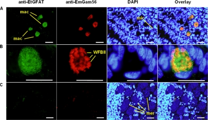
FIG. 4.
Immunolocalization of native EtGFAT in E. tenella. Paraffin sections of E. tenella-infected chicken intestine were taken at 144 h p.i. and stained with rabbit antisera developed against EtGFAT and mouse antisera developed against EmGam56. EtGFAT was detected with an FITC-conjugated anti-rabbit antibody (green) and EmGam56 with a Texas Red-labeled anti-mouse antibody (red). The sections were counterstained with DAPI (blue) for the detection of host, merozoite, and microgamete nuclei. An overlay of all three colors is shown for each image. (A) Region of tissue infected with E. tenella macrogametes (mac) and microgametes (mic); (B) closer examination of an E. tenella macrogamete; (C) region of infected tissue with E. tenella merozoites (mer). Bars, 10 μm.

