FIG. 3.
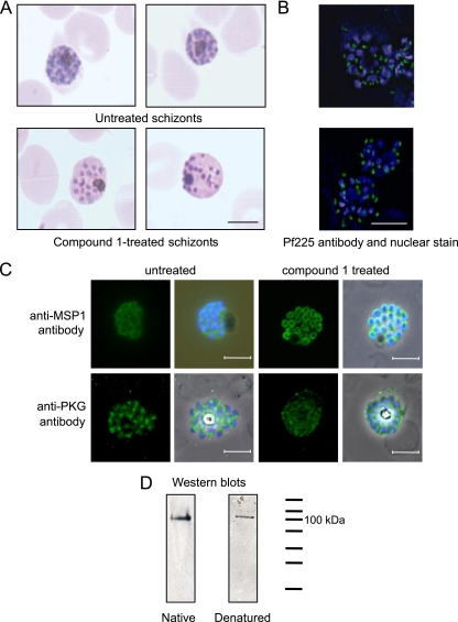
Treatment of schizonts with compound 1 causes morphological abnormalities, but merozoite proteins localize normally. (A) Giemsa-stained smears of untreated or schizonts treated with 2 μM compound 1 for 24 h. Scale bar, 5 μm. (B) Fluorescence micrographs of untreated schizonts or schizonts treated with 2 μM compound 1. The primary antibody was a monoclonal antibody that reacts with the P. falciparum rhoptry neck protein Pf225 (green). Nuclei were counterstained with 0.5 g/ml DAPI (blue). Scale bar, 5 μm. (C) Fluorescence micrographs of schizonts treated with 2 μM compound 1. The primary antibodies were a monoclonal antibody that reacts with P. falciparum MSP1 (top row) and a polyclonal antibody that reacts with PfPKG (bottom row) (both green). Nuclei were counterstained with 0.5 g/ml DAPI (blue). (D) Western blots containing mixed-stage P. falciparum (clone 3D7) parasites separated on both native and denaturing polyacrylamide gels. The PKG antibody was raised to the C-terminal peptide of human PKGI and cross-reacts with PfPKG due to the high degree of sequence identity at the C terminus.

