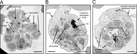
FIG. 4.
EM morphology of untreated and compound 1-treated schizonts. (A) Untreated control P. falciparum segmented schizont. Scale bar, 1 μm. (B and C) Electron micrographs showing schizonts after treatment with 2 μM compound 1 for 24 h. Hz, haemozoin; rbcm, red blood cell membrane; PVM, parasitophorous vacuolar membrane; PM, plasma membrane. Scale bar, 1 μm.

