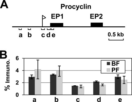
FIG. 3.
Histone H3 is less abundant on procyclin promoters in both bloodstream form and procyclic form T. brucei. (A) Schematic depicting part of an EP procyclin locus (PARP B1) (see references 62 and 67). The EP procyclin promoter is indicated with a flag, the EP procyclin genes are indicated by black boxes, and the genomic regions analyzed using qPCR are indicated by lettered brackets. (B) Histone H3 distribution over the procyclin locus was investigated using ChIP with an anti-histone H3 antibody (or no antibody as a negative control) in bloodstream form (BF) and procyclic form (PF) T. brucei. Quantitative PCR was used to amplify the procyclin genomic regions indicated in panel A. The data are expressed as the percentage of input immunoprecipitated after subtraction of signal from the no-antibody control. The results shown are the average signal from three independent ChIP experiments, with the standard deviations indicated with error bars.

