FIG. 3.
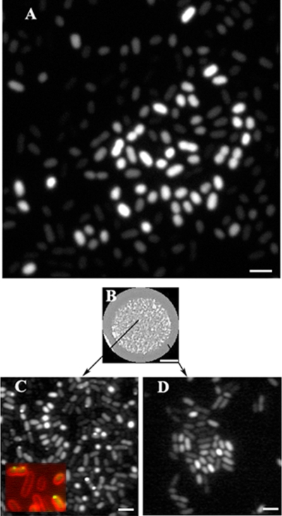
Visualization of CsgD-GFP in cells grown on agar plates. (A) Fluorescence micrograph (confocal) of CsgD-GFP-expressing strain MAE851 after 22 h of growth on LB agar without salt. Bar, 2 μm. (B) The CsgD-GFP-expressing strain MAE851 was grown as a giant “spot” colony on LB agar without salt for 72 h. Bar, 3 mm. (C and D) GFP fluorescence micrographs of bacteria that were scraped off the agar surface either from the inner (i.e., aging) parts (C) or from the outermost (i.e., young) parts (D) of the same “spot” colony. (Inset in C) Membrane localization of CsgD. Green, CsgD-GFP; red, membrane stain FM4-64. Bar, 2 μm.

