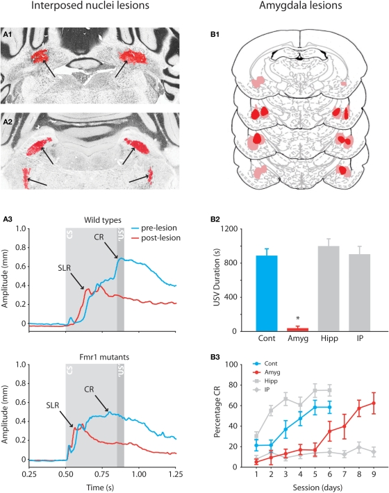
Figure 3.
Effects of anterior interposed nucleus lesions in mice and amygdala lesions in rats on eyeblink conditioning. (A1) Example of bilateral lesion (arrows) of anterior interposed nucleus in Nissl-stained section of Fmr1 mutant. (A2) Example of degenerated axonal fibers (silver staining is indicated by arrows) in the superior cerebellar peduncle and ipsilateral descending tracts. (A3) Eyeblink traces showing the average amplitudes of the CRs in wild-type and Fmr1 mutants before (blue) and after (red) the lesions. In both mutants and wild-types lesions of the anterior interposed nucleus abolish well-timed cerebellar CRs, whereas startle reflexes and SLRs are still present in the eyeblink trace. Reproduced with permission from Koekkoek et al. (2005). (B1) Histological reconstructions of amygdala lesion sites in rats. Numbers indicate distance in millimeters posterior to Bregma. (B2) Mean ultrasonic vocalization (USV) during day 6 of the training sessions. The USV duration is a valid model of anxiety in rats (Sanchez, 2003), and a reduced USV duration behaviorally confirms the lesions of the amygdala. (B3) Mean percentage of CR (±SEM) during daily training sessions from control (n = 9), interposed nuclei lesioned (n = 9), amygdala lesioned (n = 9), and hippocampus lesioned (n = 10) rats. Lesions of the amygdala robustly slowed down the acquisition of eyeblink CRs indicating that the amygdala modulates the eyeblink conditioning process. Reproduced with permission from Lee and Kim (2004).

