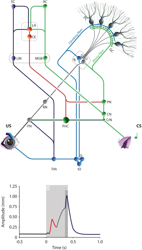
Figure 4.
The Amygdala-Cerebellum-Dynamic-Conditioning Model. An integrated model to explain the different phases in the mouse eyeblink conditioning learning process and the different peaks in the individual eyeblink traces. The colors in the model eyeblink trace represent the anatomical afferents to the FN or PnC. During eyeblink conditioning the tone (CS) and corneal air puff (US) converge at least in the amygdala and cerebellum. They are relayed to the LA from thalamic and cortical regions of the auditory (green) and somatosensory (purple) systems. Pontocerebellar (green) and olivocerebellar (blue) systems mediate the convergence of CS and US on Purkinje cells in the cerebellar cortex and to a lesser extent on the IN neurons. Amygdala and cerebellum control the FN activity via PnC (red) and RN (gray), respectively. During the first learning phase in eyeblink conditioning very efficient plasticity in the LA results in mild conditioned fear responses, including rapid facial responses such as an eyelid closure. This is represented in the eyeblink trace by an SLR (red). A few CS-US pairings are enough to obtain clear SLRs in mice. Prolonged training will induce cerebellar learning, which behaviorally is represented by a perfectly timed eyelid closure (gray). In addition, direct projections from the amygdala to the PN might contribute directly to the CS input of the cerebellum. Thus, the ACDC model assumes that neuronal mechanism in different brain regions contribute to the establishment of an adequate CR. AC, Auditory cortex; CE, Central amygdala; CN, Cochlear nucleus; CrN, Cochlear root nucleus; CS, Conditioned stimulus; FN, Facial nucleus; GC, Granule cell; IN, Interposed nuclei; IO, Inferior olive; LA, Lateral amygdala; MGB, Medial geniculate body of the thalamus; MN, Motor neurons; PC, Purkinje cell; PIN, Posterior intralaminar nucleus of the thalamus; PN, Pontine nuclei; PnC, Caudal pontine reticular nucleus; RN, Red nucleus; SC, Somatosensory cortex; TrN, Trigeminal nerve nucleus; US, Unconditioned stimulus.

