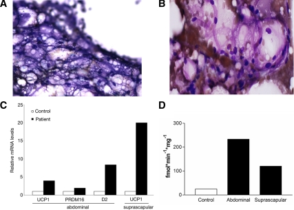
Figure 2.
Histological and molecular studies on adipose from the patient with type A insulin resistance. A, Dif-quick staining at 120× magnification of BAT from the suprascapular needle biopsy showing multivesicular cells containing lipid. B, Hematoxylin and eosin staining at 200× magnification of adipose tissue from periumbilical sc abdominal needle biopsy showing an atypical appearance consistent with brown adipose. The adipocytes lacked features of white adipose (large empty cell with classical signet-ring morphology). C, Molecular signature of brown fat in the adipose depots of the patient: real-time PCR analysis of mRNA levels of the brown fat molecular markers UCP1, PRDM16, and D2 in abdominal sc fat tissue and of UCP1 in suprascapular fat depot in a control subject and the patient. D, Type 2 5′ deiodinase activity in adipose biopsies obtained from the patient’s suprascapular and sc abdominal depots compared with control sc abdominal adipose biopsy. The patient’s type 2 5′ deiodinase activity is higher in both abdominal (white adipose) and suprascapular (brown adipose) regions compared with control.

