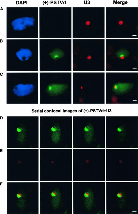
Figure 4.
Asymmetrical Distribution of U3 snoRNA in the Presence of the (+)-PSTVd within the Nucleolus.
(A) Symmetrical distribution of U3 in the nucleolus of a mock-inoculated plant.
(B) and (C) Two infected cells showing the asymmetrical distribution of U3 (red) and PSTVd (green) in the nucleoli. Note that in each case the two RNAs occupy separate domains within the nucleolus with little overlapping.
(D) to (F) Serial confocal images showing the asymmetrical distribution of PSTVd (green in [D]) and U3 (red in [E]). The merged images ([F]) show little overlapping of the two RNAs.
Bars = 2 μm.

